
Normal Cervical MRI
Quick Summary
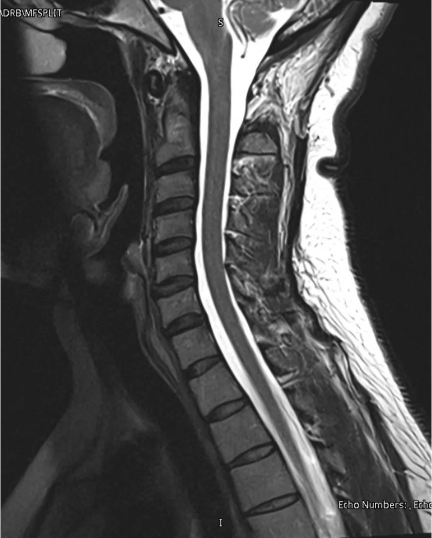
A normal cervical MRI shows healthy discs, well-aligned bones, and open spaces for the spinal cord and nerves. There is no significant cord compression, nerve compression, or structural damage.
Understanding a Normal Cervical MRI
1. Bones, Discs, and Alignment
A cervical MRI shows the neck bones (vertebrae) stacked on top of each other with smooth alignment. Between each bone is a disc, which acts as a cushion and allows the neck to bend and rotate. On a normal MRI, the discs appear well hydrated and maintain their height.
The spinal canal, which protects the spinal cord, appears open with adequate space.
2. Spinal Cord and Nerves
The spinal cord runs through the center of the neck, carrying signals between the brain and the rest of the body. On a normal MRI, the cord appears round and well-cushioned, without flattening or compression from discs, bone spurs, or thickened ligaments.
Nerves branch off the spinal cord and exit through small openings called foramina, traveling into the shoulders, arms, and hands. On a normal MRI, these nerves have enough room and are not pinched.
Healthy cord and nerve function is important for normal strength, sensation, and coordination in the arms and hands, as well as balance and walking.
3. Soft Tissues and Supporting Structures
Ligaments, joints, and muscles help support the neck. On a normal MRI, these structures appear intact without significant inflammation or injury.
A normal cervical MRI does not show disc herniation, spinal stenosis, myelopathy, fractures, or instability.

When should I see a spine specialist?
You should seek evaluation if you have:
Neck pain lasting more than several weeks
Arm pain, numbness, or weakness
Hand clumsiness or difficulty with fine motor tasks
Balance problems or difficulty walking
Symptoms that interfere with daily activities
Early evaluation can help guide appropriate treatment.